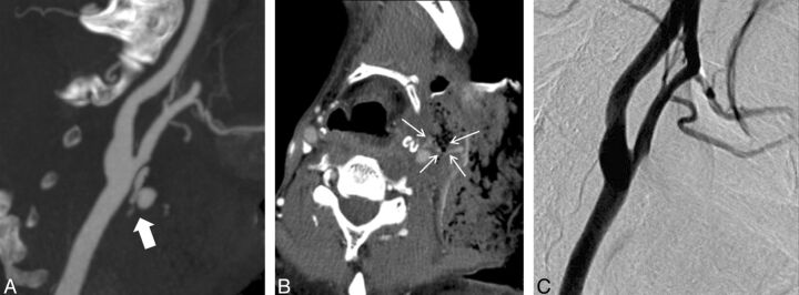
Fig 1.
A 52-year-old man with a history of oropharyngeal cancer. A, MIP CTA imaging performed after bleeding was controlled by local packing shows contrast extravasation (arrow) from the common carotid artery near the bifurcation. B, Source image shows an exposed common carotid artery (arrows) surrounded by necrosis. C, DSA done immediately after CTA does not show contrast extravasation. However, the possibility of a further tamponade effect after CTA cannot be excluded in this case.

