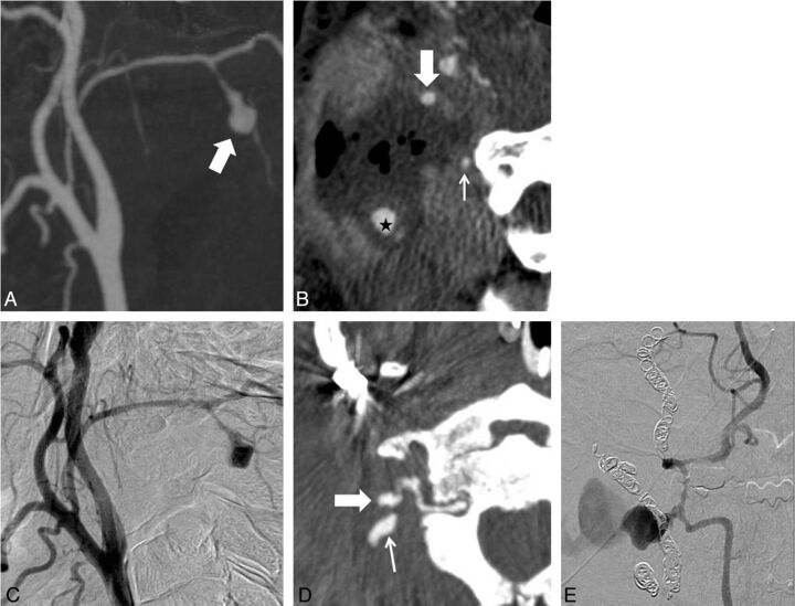
Fig 2.
A 42-year-old man with a history of nasopharyngeal cancer. A, MIP CTA image shows a pseudoaneurysm (arrow) at the right occipital artery. B, Oblique reformatted image shows the pseudoaneurysm surrounded by necrotic tissue (star). Half of the circumference of the right internal carotid artery (large arrow) is exposed to the necrotic tissue, and the unexposed vertebral artery (small arrow) is close to the necrotic margin. C, DSA confirms the pseudoaneurysm of the occipital artery. The pseudoaneurysm is embolized with coils. At 64 days after treatment, another blowout occurs. D, Oblique reformatted CTA image shows a newly developed pseudoaneurysm (large arrow) with extravasation from the right vertebral artery (small arrow). E, DSA confirms the diagnosis.

