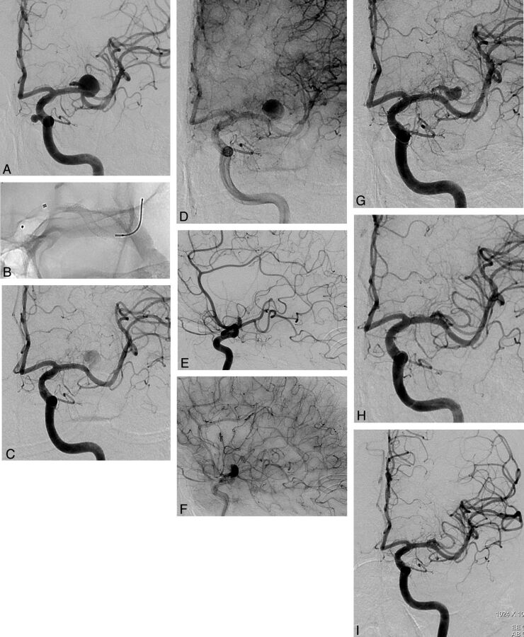
Fig 2.
Occlusion stages of left MCA bifurcation aneurysm. A, DSA image shows the aneurysm giving rise to superior trunk. B, Fluoroscopic image shows the deployment of the PED in the inferior trunk. C and D, Early and late phases of 6-month control angiogram demonstrate the reduced and delayed filling of the aneurysm sac with the significant stagnation. Bifurcating branch is also filling belated in reduced caliber. E and F, Early and late phases of 6-month control angiogram (lateral view) show reduced filling of the superior trunk with retrograde filling of the distal branches through pial collaterals. G, One-year control angiogram demonstrates the remodeled superior trunk. The superior trunk and its branches are still filling in reduced caliber. H, Eighteen-month control angiogram shows complete occlusion of the aneurysm, with the superior trunk coming to its original size. I, Thirty-month control angiogram shows the stable occlusion of the aneurysm with the patency of the bifurcating branch (note the carotid cave aneurysm in A, treated with PED as well).

