
Fig 2.
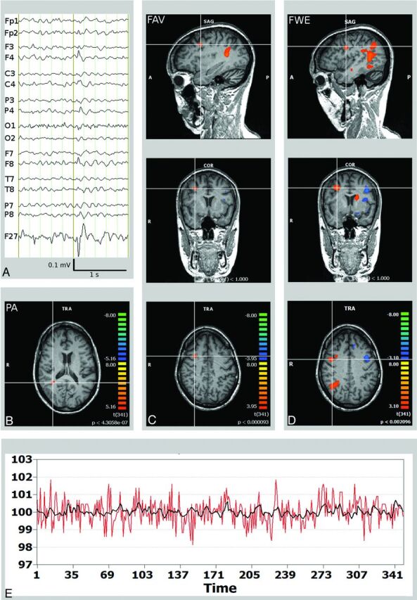
Right frontal lobe epilepsy and frontal focal cortical dysplasia (patient 12). A, Scalp EEG (average reference montage) and IC-factor coding for the interictal spike with right frontal maximum. B, Misleading localization of the PA in the right inferior parietal lobule (crosshair). C, FAV delineates a BOLD signal cluster matching the right frontal focal cortical dysplasia/SOZ (crosshair). D, The widespread BOLD signal correlates obtained by the FWE were not of use in planning of invasive EEG recordings. E, BOLD time course of the right frontal SOZ (red, in a percentage of signal change). Time course of the independent component factor derived from the EEG data after convolution with the hemodynamic response function representing the epileptic activity (black).