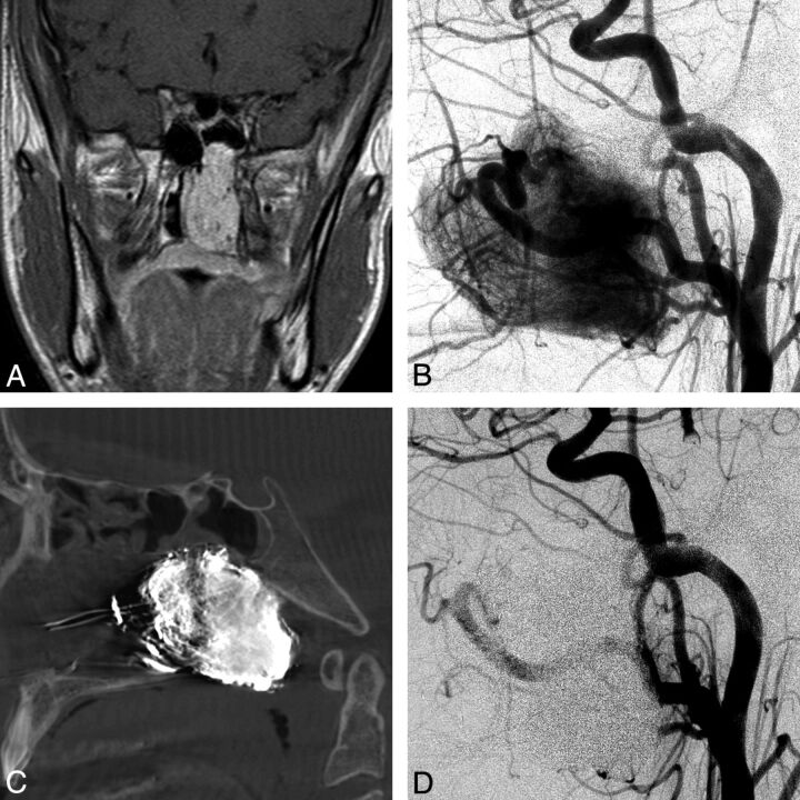
Fig 2.
A 13-year-old boy with nasal stuffiness and a Fisch Ib juvenile angiofibroma. A, Coronal contrast-enhanced T1-weighted MR image shows a contrast-enhancing mass within the left nasal cavity and posterior nasopharynx, extending into the left sphenoid sinus. B, Lateral left common carotid angiogram (LCCA) shows the hypervascular mass with supply off the ECA and ICA branches. C, Lateral DynaCT (Siemens, Erlangen, Germany) image shows the EVOH (Onyx) cast within the tumor. D, Lateral LCCA postembolization shows no filling of the hypervascular mass.

