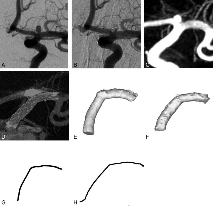
Fig 1.
Illustrative case of a 61-year-old female with family history of aneurysmal subarachnoid hemorrhage. MR angiography revealed a left ICA terminus aneurysm. Catheter angiography demonstrated a 2.5 mm aneurysm having a 2 mm neck (A, frontal oblique projection) that subsequently underwent stent assisted coil embolization (B, frontal oblique projection). Pre-embolization 3DRA (C) and post-embolization CBCT (D) are used to isolate a 3D model of the stented artery (pre-embolization, E; post-embolization, F). The centerlines of the stented vascular segment pre and post stent-assisted coiling are extracted in G and H, respectively.

