Fig 1.
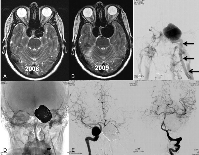
62-year-old woman with incidentally discovered bilateral cavernous sinus aneurysms. A, MR imaging for headaches in 2006 shows bilateral cavernous sinus aneurysms. Treatment was not recommended. B, MR imaging 3 years later demonstrates growth of both aneurysms. The patient was asymptomatic. C, In 2010, the left aneurysm ruptured causing a carotid cavernous fistula with proptosis and ophthalmoplegia. Left carotid angiogram in frontal view shows early filling of both cavernous sinus and many skull base and neck veins. There was no filling of intracranial vessels. Arrows indicate internal carotid artery. D, The aneurysm was coiled; the carotid artery was also occluded with coils and sealed with a balloon. Clinical symptoms were cured within 2 weeks. E and F, Frontal views of right internal carotid artery (E) and left vertebral artery (F) after left internal carotid artery occlusion demonstrate collateral flow to the left hemisphere via the circle of Willis.

