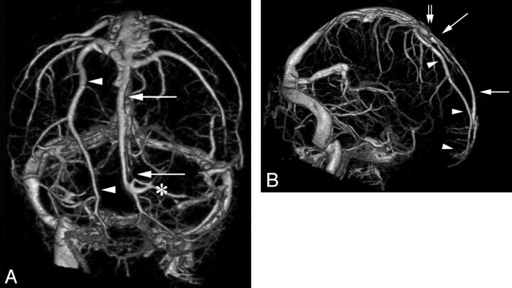
Fig 1.
CTA, venous phase, obtained with a 320-MDCT in a 41-year-old woman imaged for pseudotumor cerebri. Anteroposterior (A), and right lateral (B) projections of the subtracted intracranial venous system. Unilateral hypoplastic rostral SSS is documented on the right side. Note the compensatory drainage through an enlarged parasagittal frontal cortical vein (arrowhead), coursing parallel to the midline from the pole of the right frontal lobe to the SSS in the region of the coronal suture (double arrow). A hemi-SSS (arrows) is observed on the left side, directly continuous with the middle third of the SSS. Left frontopolar cortical veins (asterisk) drain into the most rostral portion of the SSS at a right angle.

