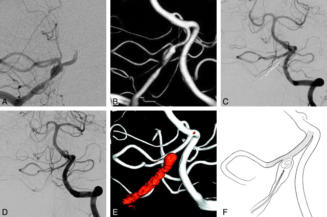
Fig 1.
A and B, The anteroposterior view of the right vertebral artery angiography (A) and the 3D reconstruction image (B). C, The Enterprise stent is placed through the PICA to the distal portion of right vertebral artery via left the vertebral artery approach. D, The 6-month follow-up angiography reveals good PICA patency. E, The 3D reconstruction image also shows good PICA patency. F, Illustration of the vertebral artery-to-PICA stent placement with coil embolization.

