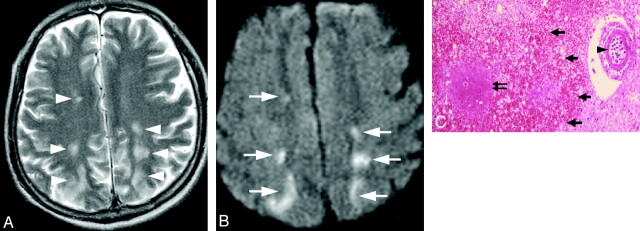
Fig 1.
Case 1. Infarctlike lesion in a 74-year-old man. A, Axial T2WI shows hyperintense spots in the bilateral watershed area (white arrowheads). B, The corresponding areas show diffusion restriction on DWI (white arrows). C, Pathologic specimen shows irregular-shaped necrosis (double black arrow) and congestion (thick black arrows) surrounding the occluded vessel (arrowhead) (hematoxylin-eosin, original magnification ×25).

