Fig 3.
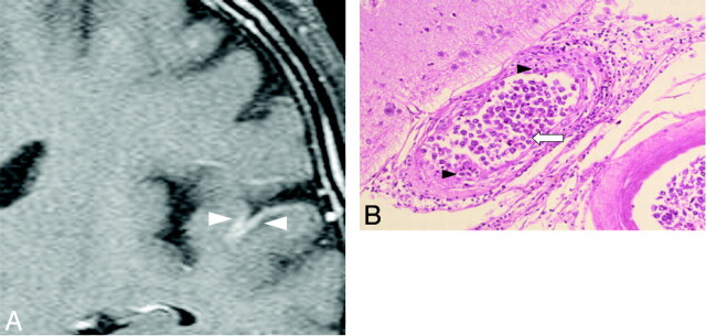
Case 2. Meningeal enhancement in a 69-year-old man. A, Gadolinium-enhanced coronal T1-weighted image shows abnormal meningeal enhancement around the temporal lobe before treatment (white arrowheads). B, Pathologic specimen shows thickening of the affected vessel walls with intraluminal (white arrow) and subendothelial (black arrowheads) tumoral infiltration (hematoxylin-eosin, original magnification ×25).

