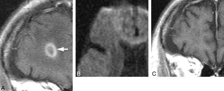
Fig 4.
Case 4. Masslike lesion in a 70-year-old man. A, Coronal gadolinium-enhanced T1-weighted image shows ringlike enhancement before treatment (white arrow). B, Axial DWI shows no abnormal signal intensity in the lesion. C, Follow-up gadolinium-enhanced coronal T1-weighted image shows regression of the enhancement after treatment on day 121.

