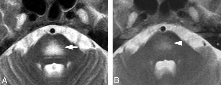
Fig 5.
Case 8. Hyperintense lesion in the pons on a T2-weighted image in a 63-year-old woman. A, Axial T2WI shows symmetric hyperintense lesions in the center of the pons sparing the pontine tegmentum and ventrolateral pons (white arrow). DWI showed T2 shinethrough without diffusion restriction (not shown). B, T2WI after chemotherapy shows a decreased abnormal signal intensity in the pons on day 85 (white arrowhead).

