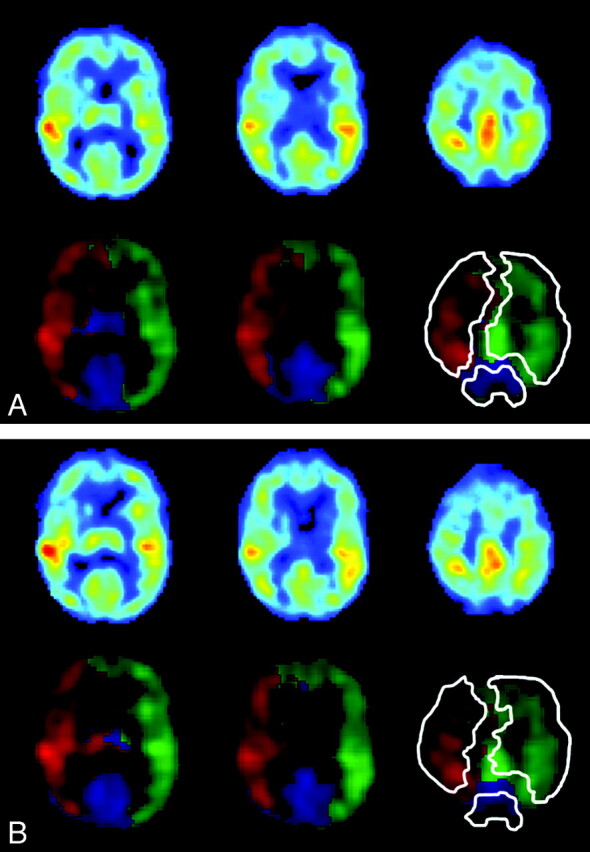
Fig 3.

Three axial sections showing perfusion-weighted images, flow territories, and manual outlining of the ICAs and the BA in an 80-year-old female patient with right ICA stenosis of 70%–99%. A and B, Images are based on the first (A) and last (B) 35 dynamics.
