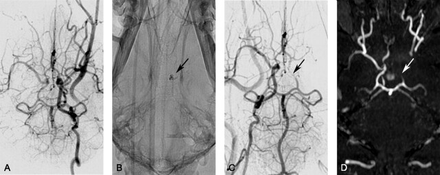
Fig 1.
Representative of DSA and TOF imaging canine stroke modeling: angiographic data of a single subject obtained during embolic stroke induction. A, DSA of the left ICA before introducing the autologous clot (ventral projection). B, The injected blood clot is seen on radiography due to the presence of barium sulfate (arrow). C and D, DSA (C, ventral projection) and 3D time-of-flight image (D, ventral projection) of the right ICA confirm occlusion of the distal left ICA and proximal MCA (arrows).

