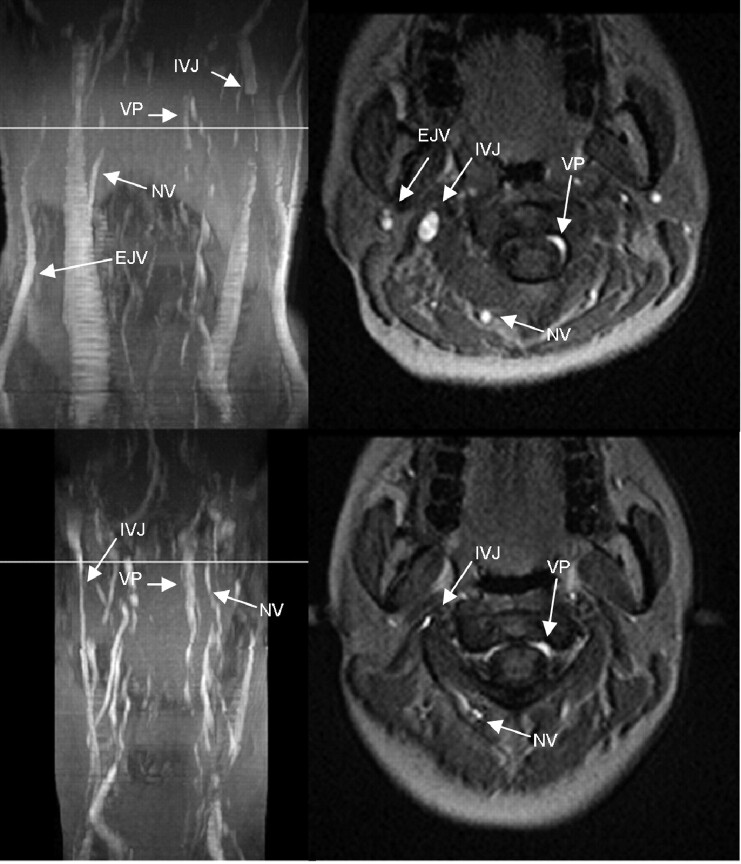
Fig 4.
MIP (left column) and axial source images at the level of C2 in supine (top row) and sitting positions (bottom row) in a healthy volunteer. In the supine position, there is a high-grade stricture of the left IJV. The EJV, NV, and VP are visible. In the sitting position, only the right IJV is visible, but narrowed. The VP are prominent, and the NV can be identified.

