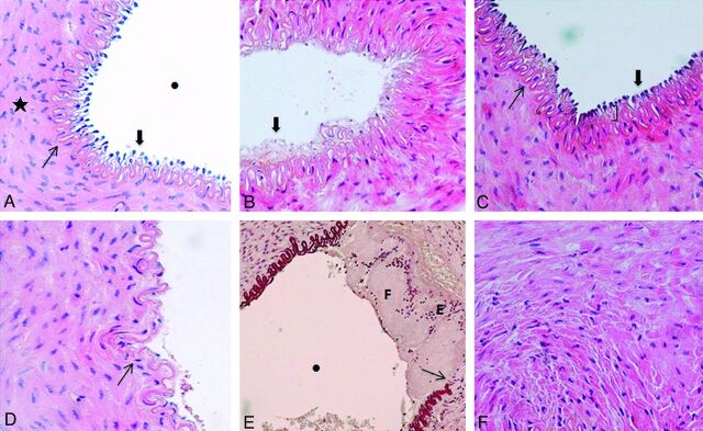
Fig 3.
Histopathologic grading systems. A, Microscopic view of a right SFA (control sample) (hematoxylin-eosin [HE] staining, original magnification × 400) demonstrates well-maintained tissue integrity throughout the internal layers of the vessel. Endothelial cells (thick arrow) protruded toward the vascular lumen (dot), and the IEL (thin arrow) is intact. The mediaI layer (star) has homogeneous layers of smooth muscle cells. B, Microscopic view of a left SFA (device sample) (HE staining, original magnification × 400) shows total denudation of the endothelium (thick arrow) (100% of the surface area). C, Microscopic view of a left SFA (device sample) (HE staining, original magnification × 400) shows focal intimal thickening (parentheses) due to edema. Endothelial cells (thick arrow) and the IEL (thin arrow) are intact. D, Microscopic view of a left SFA (device sample) (HE staining, original magnification × 400) shows a focal fracture (thin arrow) of the IEL and denudation of the endothelium. E, Microscopic view of a left SFA (device sample) (orcein staining, original magnification × 200) demonstrates a fracture of the IEL (thin arrow) and mural thrombus with a layered pattern including both fibrin-rich (F) and erythrocyte-rich (E) layers. The mural thrombus does not compromise the lumen of the vessel (dot). F, Microscopic view of a left SFA (device sample) (HE staining, original magnification × 400) demonstrates edema within the media layer visualized as interstitial infiltration among smooth-muscle cells.

