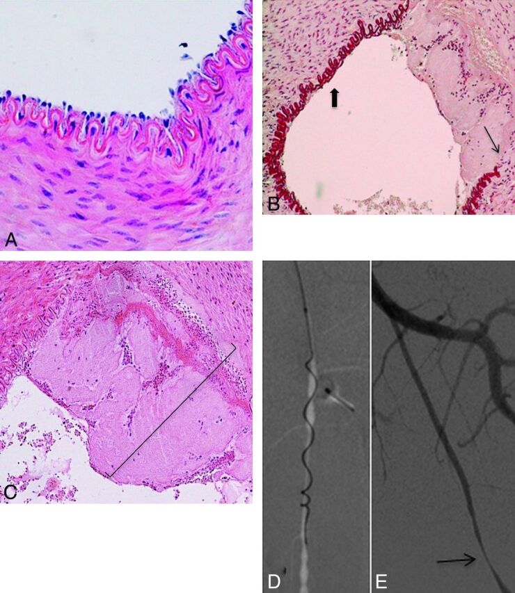
Fig 5.

Histopathologic findings after mechanical thrombectomy by using the Catch thromboembolectomy system. A, Microscopic view of a right SFA (control sample) shows all layers preserved (hematoxylin-eosin [HE] staining, original magnification × 400). B, Microscopic view of a left SFA demonstrates the denudation of endothelial cells (thick arrow) and the broken IEL (thin arrow) (orcein staining, original magnification × 200). C, Microscopic view of a left SFA shows a mural clot (parentheses) and the IEL fractured. (HE staining, original magnification × 200). D, Anteroposterior angiogram of the left SFA shows the Merci clot device during the retrieval. E, Anteroposterior angiogram of the left SFA immediately after thrombectomy demonstrates focal stenosis (thin arrow).
