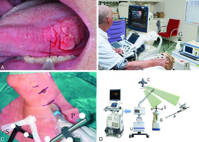
Fig 1.
Patient 2 with a T2 lateral tongue carcinoma. A, Peritumoral injection of technetium Tc99m labeled–nanocolloid. B, Freehand SPECT-US. Note the optical markers on the freehand gamma camera (G), the transmitter generating the magnetic field (T), the electromagnetic sensor attached to the US transducer (Tr), and the shared optical and electromagnetic sensor (S) affixed to the head of the patient. The optical camera is not shown in this image. A fused SPECT-US image is on the screen (Sc) after SPECT data have been loaded onto the US machine in DICOM format by using a USB stick. C, Use of freehand SPECT during surgery. Note the optical sensors on the probe (P) and the sternum of the patient (S). D, Freehand SPECT and US system with infrared optical tracking system (camera [C], detectors [G+P], and sensors [S]) and a data processing unit (U).

