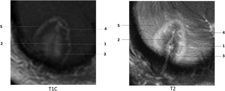
Fig 2.
Concentric zones: T1 contrast-enhanced (T1C) and T2WI 24 hours after laser ablation of a recurrent right cerebellar metastatic lesion in a 71-year-old female patient with history of breast carcinoma: 1) probe track, 2) central zone, 3) peripheral zone, 4) peripherally enhancing rim, 5) marginal zone. Note that the concentric zones appear as inverse images on T1C and T2 images.

