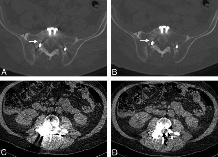
Fig 4.
A 65-year-old man status post L3-to-S1 pedicle screw and rod fixation. wFBP (A) and IMAR (B) images at the S1 level using bone window settings demonstrate lucency about the right S1 screw, consistent with hardware loosening (white arrows). At the L3 level, the central canal and lateral recesses are obscured by artifacts on wFBP (C) image with soft-tissue window settings. IMAR (D) image with soft-tissue window settings at the same level demonstrates a retained wire from a prior spinal cord stimulator (wavy arrow) and improved visualization of the left lateral recess (black arrowheads).

