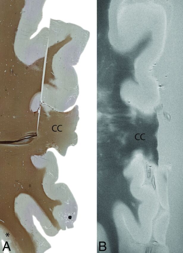
Fig 2.

An example of extensive cortical demyelination in an MS case. Histologic section with anti-proteolipid protein antibody (left) and a matched T2WI (right). The histologic section shows extensive cortical demyelination (lack of proteolipid protein) in the cortex, except for a small section at the left bottom (asterisk). This extensive demyelination makes it difficult to differentiate lesions and normal-appearing gray matter on MR imaging (right). In this particular case, as a result, prospective MR imaging scoring was negative. CC indicates corpus callosum. Degree of magnification: 50×.
