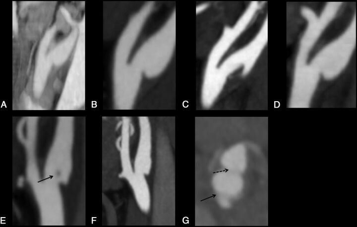
Fig 1.
Sagittal and axial images of carotid webs. The top panel shows serial sagittal-view CTAs in patient A. A and B, Carotid webs 8 years apart. C, Changing morphology of the right carotid web within days with possible thrombus formation in the setting of dual antiplatelet therapy. D, A return to baseline morphology with the use of unfractionated heparin. The bottom panel shows serial CTAs in patient B. E, A carotid web with possible thrombus in the lumen (arrow). F, The same carotid web 1 month later with no thrombus. G, An axial-view CTA with the same carotid web appearing as a septum (arrow). The broken arrow indicates the carotid bifurcation.

