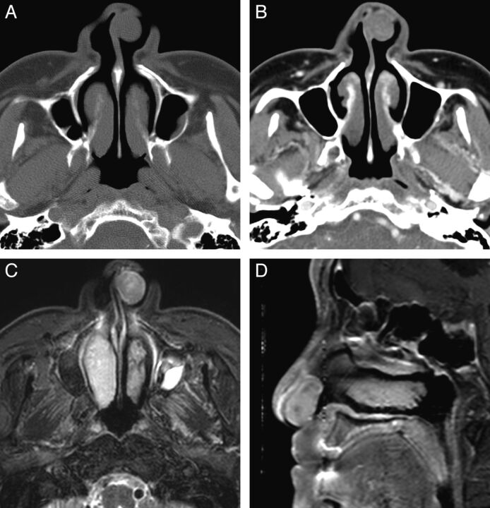
Fig. 1.
Case 10. Schwannoma of the nasal cavity in a 79-year-old man. A, Precontrast axial CT scan with a bone algorithm shows a polypoid mass in the left anterior nasal cavity, originating from the nasal septum. B, Contrast-enhanced axial CT scan with a soft-tissue algorithm shows mild and patchy enhancement of the mass. C, Fat-suppressed axial T2-weighted MR image shows that the mass is isointense to the brain stem. D, Contrast-enhanced fat-suppressed sagittal T1-weighted MR image shows marked contrast enhancement within the mass.

