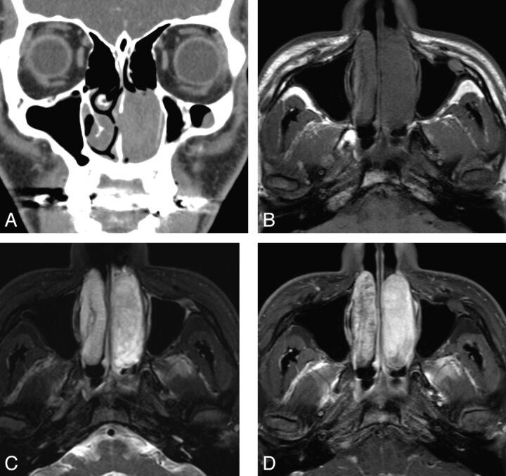
Fig. 2.
Case 5. Schwannoma of the nasal cavity in a 22-year-old man. A, Contrast-enhanced coronal CT image shows a tubular expansile soft-tissue mass in the left nasal cavity, demonstrating mild enhancement. B, Axial T1-weighted MR image shows the tumor extending in the anteroposterior dimension and remodeling the lateral nasal wall. C, Fat-suppressed axial T2-weighted MR image shows that the tumor is hyperintense to the brain stem. D, Contrast-enhanced fat-suppressed axial T1-weighted MR images show marked homogeneous enhancement within the tumor.

