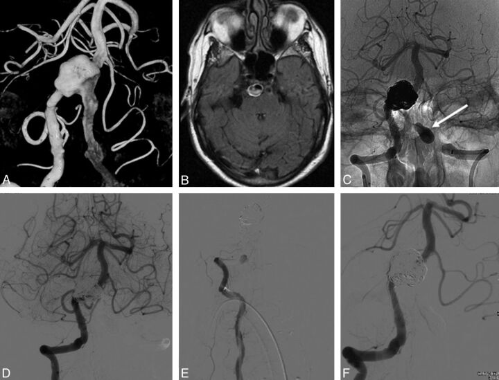
Fig. 5.
A 64-year-old man with abducens paresis (patient 7). A and B, 3D angiography and MR imaging demonstrate a large dolichoectatic partially thrombosed basilar trunk aneurysm. Note irregular and ectatic distal vertebral arteries. C and D, Right vertebral angiography after telescopic placement of 2 LEO stents and a Silk flow diverter, coiling of the aneurysm lumen, and balloon occlusion of the left vertebral artery in the V4 segment (arrow in C). E, After embolization, the patient did not wake up from general anesthesia. Repeated right vertebral angiography shows in-stent thrombosis in the V4 segment. F, Recanalization of the basilar artery after mechanical thrombectomy but with persistent occlusion of the right posterior cerebral artery. The patient died the same day.

