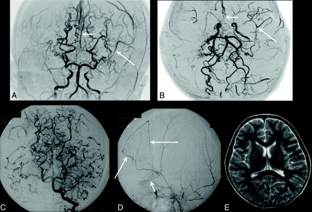
Fig 3.
A 7-year-old girl (diagnostic age group, 4–7 years) who had recurrent transient left hemiparesis for 5 years was diagnosed with Moyamoya disease, which manifested clinically as TIA. A and B, Coronal and axial time-of-flight MR angiograms show advanced steno-occlusive changes at or around the terminal part of the right ICA, with poorly visualized ACA and MCA branches (ICA stage III, right). Moderate steno-occlusive changes at or around the terminal part of the left ICA with relatively good visualization of the ACA (small arrows) and MCA (large arrows) cortical branches (ICA stage II) are seen on the left. No steno-occlusive lesions are seen bilaterally in the posterior cerebral artery (PCA stage 1, bilaterally). C, Anteroposterior view of the vertebral angiogram shows no steno-occlusive lesions bilaterally in the PCA and well-developed leptomeningial collateral circulation to the anterior circulation. D, Left lateral external carotid angiogram shows dilated anterior branches of the middle meningeal artery providing transdural collaterals to the contralateral frontal region (large arrows), with the medial branches of the maxillary artery providing transdural collaterals to the right anterior basal region (small arrow). Two transdural collaterals can be seen on the right, and none are seen on the left (anteroposterior view of the left external carotid angiogram, not shown). E, Axial T2-weighted MR image shows no infarction bilaterally (ie, the number of infarcted regions is zero on either side). Note that in this patient, the steno-occlusive changes do not involve the PCA, even in the right hemisphere, where ICA lesions are advanced (stage III).

