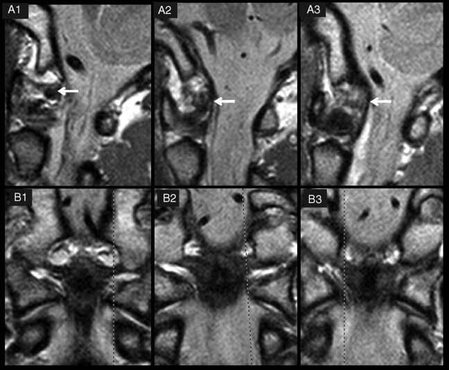
Fig 4.
Three examples of grades of delineation and signal intensity of alar ligaments. A and B, PD−weighted MR images of 3 alar ligaments in the sagittal (A) and coronal (B) planes. The dashed lines in B1–3 indicate the orientation of the respective sagittal sections in A1–3. In A1, the alar ligament shows a good delineation (grade 3) and low signal intensity (grade 0). A2 is classified as grade 2 for signal intensity (high signal intensity in one-third to two-thirds of the cross-section) and delineation (moderate). In A3, the alar ligament is hardly definable (grade 1), with high signal intensity throughout the cross-section (grade 3).

