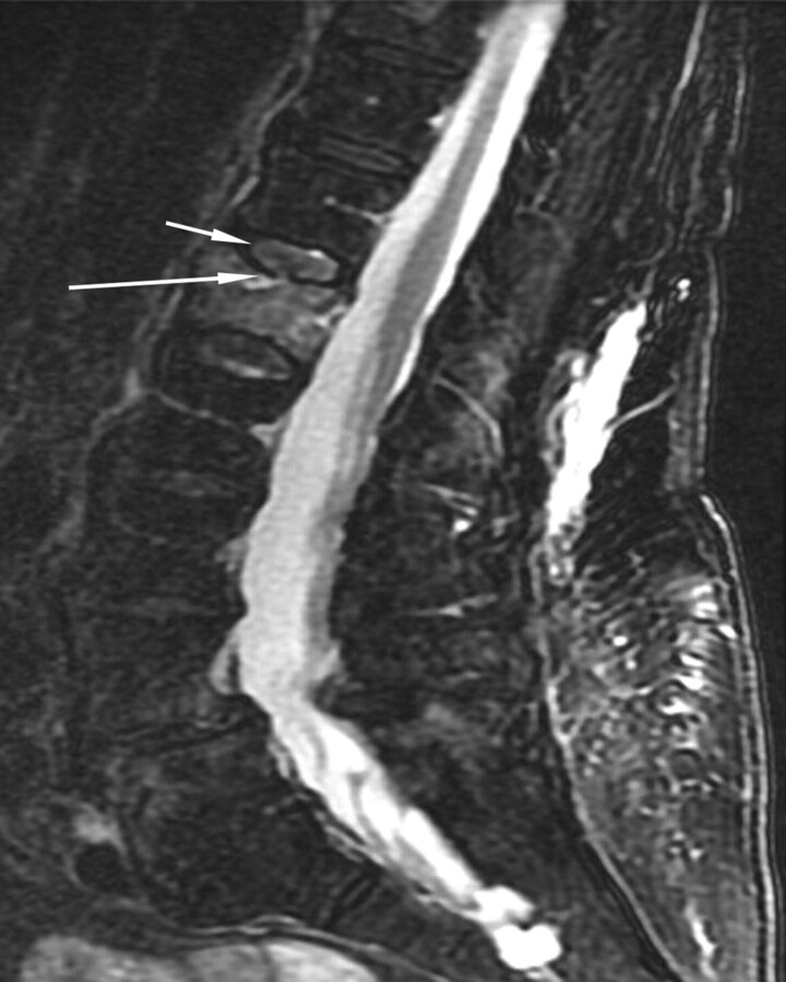
Fig 1.
An 88-year-old woman with a 3-week history of severe low back pain. Fat-suppressed T2-weighted sagittal MR image shows diffuse marrow edema within the L1 vertebra and mild height loss consistent with an acute osteoporotic vertebral compression fracture. Focal cortical discontinuity (large arrow) is seen within the superior endplate due to a fracture. This is associated with abnormally increased signal intensity within the T12-L1 intervertebral disk (small arrow), which is consistent with injury to this structure.

