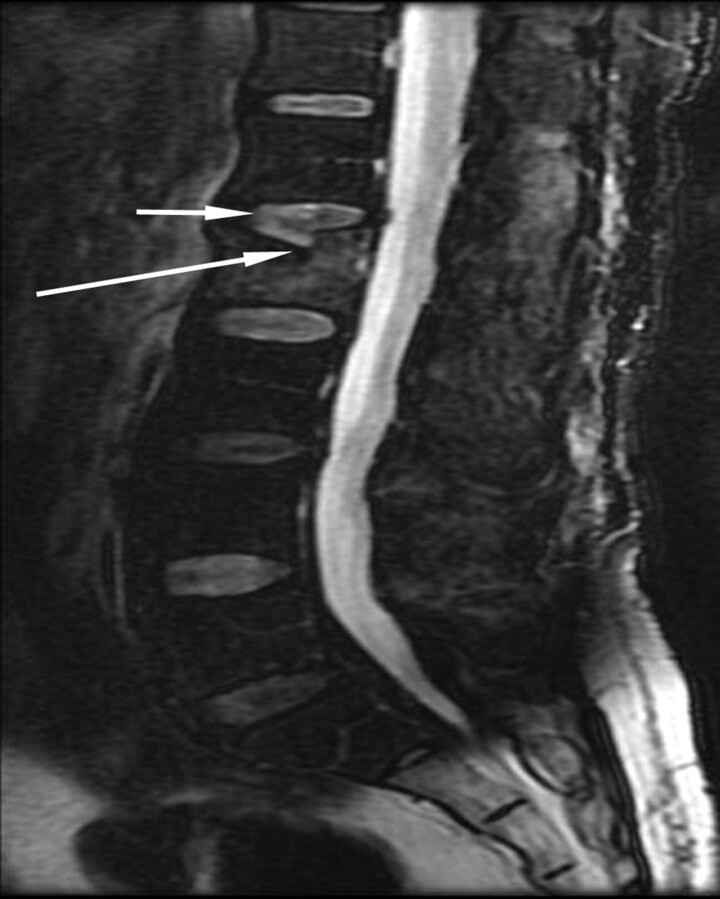
Fig 2.
A 62-year-old man with diabetes admitted with a 1-week history of debilitating low back pain. Fat-suppressed T2-weighted sagittal MR image shows diffuse marrow edema within the L2 vertebra. Inferior angulation of a fractured superior endplate (long arrow) is observed. Note the alteration of signal-intensity change and morphology of the injured L1–2 intervertebral disk (small arrow) compared with the neighboring intervertebral disks.

