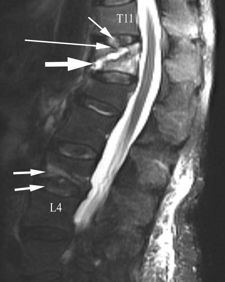
Fig 3.
A 79-year-old woman with a 6-week history of low back pain. Fat-suppressed T2-weighted sagittal MR image shows diffuse marrow edema within the T12 vertebra and mild height loss consistent with an acute osteoporotic vertebral compression fracture. A large fluid-filled fracture line (bold arrow) is seen coursing obliquely toward a fractured superior endplate (large arrow). The T11–12 intervertebral disk demonstrates edema (small arrow). An inferior L3 endplate fracture is associated with L3–4 intervertebral disk edema (double arrows). A chronic L1 vertebral compression deformity is present.

