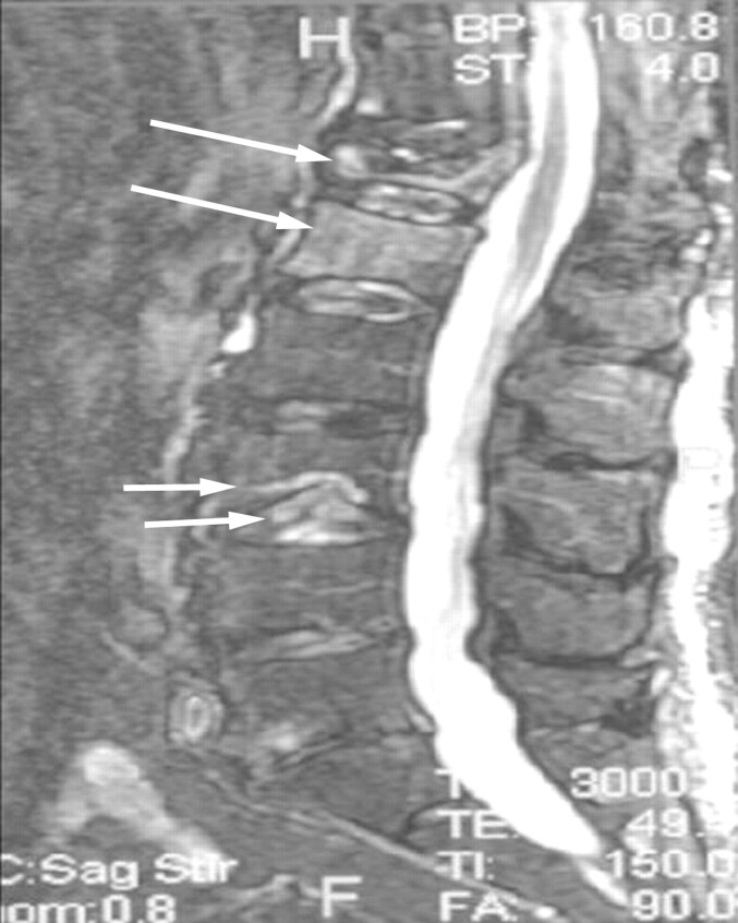
Fig 6.
An 82-year-old man with a 3-week history of severe progressive low back pain. Fat-suppressed T2-weighted sagittal MR image shows diffuse marrow edema within the L1 and L2 vertebrae (large arrows), consistent with acute osteoporotic vertebral compression fractures. The superior endplate of T12 is fractured, and the adjacent disk shows foci of edematous change. An inferior L3 endplate fracture is associated with injury to the adjacent intervertebral disk (double arrows). These latter findings were not reported.

