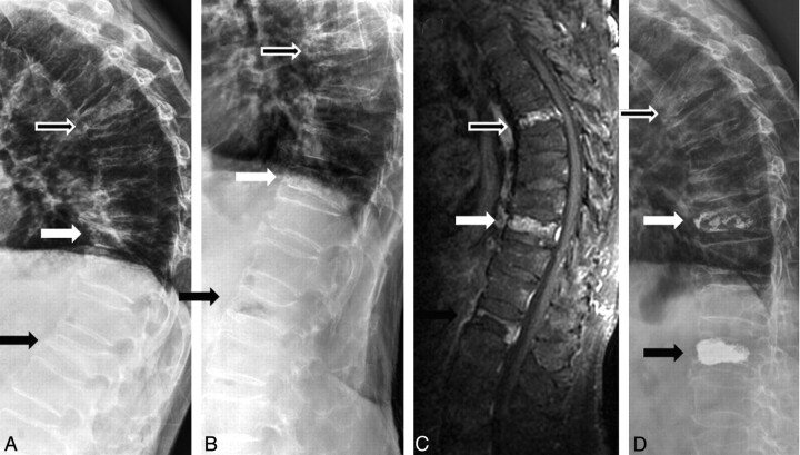
Fig 3.
A 74-year-old man with severe back pain due to T6, T10, and L1 compression fractures. A and B, Sitting (A) and supine with bolster (B) lateral radiographs show mobility at L1 (black arrow) but no mobility at T6 (hollow arrow) and T10 (white arrow). C, Contrast-enhanced T1-weighted MR image demonstrates contrast enhancement at T6, T10, and L1. D, Postvertebroplasty lateral radiograph shows cement filling in T10 and L1. Vertebroplasty was not performed at T6, but the patient still showed dramatic improvement.

