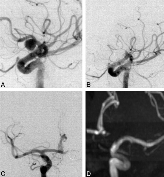
Fig 1.
A and B, Left carotid DSA oblique views show the aneurysm on the proximal third of the left A1 segment (A) and the result immediately after EVT (B). C, Right carotid DSA, anteroposterior view, after EVT shows the patency of the AcomA and contralateral A1 segment with filling of the distal left A1 segment. D, Maximum-intensity-projection reconstruction of the left carotid artery on an MRA 2.5 years after EVT.

