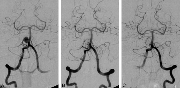
Fig 2.
A 72-year-old woman with a proximal AICA aneurysm, which originated from the AICA-PICA variant. A, Right VA angiogram reveals a wide-neck saccular aneurysm on the proximal right AICA-PICA variant. B, The right VA angiogram confirms partial occlusion of the aneurysm with preservation of the AICA-PICA variant immediately after embolization. C, The follow-up angiogram shows residual neck filling of the aneurysm with minimal coil compaction 12 months later.

