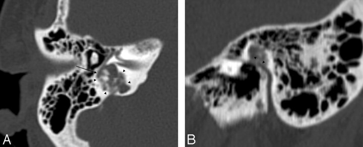
Fig 1.
A, Axial CT scan shows an osteolytic heterogeneous mass, with central calcifications invading the posterior labyrinth (arrowheads). Note the close relationship between the second portion of the facial nerve and the tumor (arrow). B, Sagittal CT scan reformat shows a close relationship (arrowheads) between the tympanic segment of the facial nerve and the tumor.

