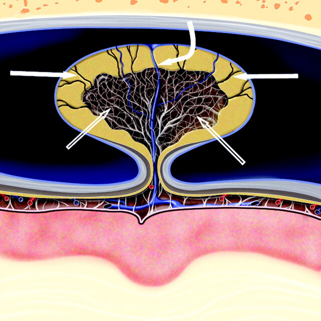
Fig 4.
Cross-sectional graphic of a giant venous sinus AG projecting into a dural venous sinus. A core of CSF-filled collagenous trabeculation (open arrows) extends from the subarachnoid space into the granulation and is covered by an apical cap of arachnoid cells. CSF channels (arrows) extend through the cap to the sinus endothelium and drain CSF into the venous circulation. A vein (curved arrow) also courses through the body of the AG, penetrates the arachnoid cap layer, and empties into the dural venous sinus. Graphic is used with permission from Amirsys Inc., Salt Lake City, Utah

