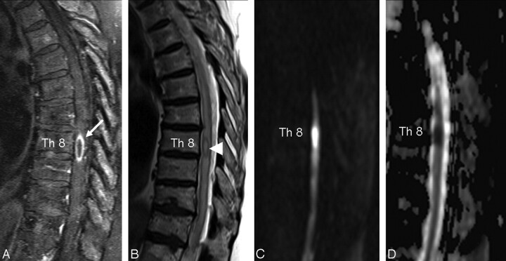
Fig 1.
A, Sagittal fat-saturated T1-weighted turbo spin-echo sequence after intravenous Gd-chelates application shows an intramedullary ring-enhancing lesion at T8 (white arrow). B, Sagittal T2-weighted turbo spin-echo sequence demonstrates an extended thoracic spinal cord edema and a hypointense capsular rim of the lesion (white arrowhead). C, The lesion is hyperintense on sagittal DWI. D, Restricted diffusion is illustrated on ADC maps.

