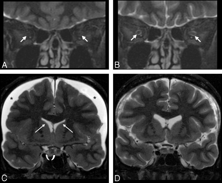
Fig 2.
Patient sample 1. Cranial MR imaging (coronal STIR sequence) of a 49-year-old woman (patient 6) with CSH due to spontaneous CSF leak in the spinal dura before (A and C) and on follow-up imaging after (B and D) successful therapy with an epidural blood patch. Imaging of the orbital contents reveals a lack of the normal peri-optical CSF rim in the ISSON (arrows in A, collapsed ONS), which normalized with therapy (arrows in B). Width of the ISSON is 1.3 mm (position 1), 1.3 mm (position 2), 1.0 mm (position 3), and 0.9 mm (position 4) on follow-up. Lower normal width of the ISSON is 1 mm (position 1) and 0.9 mm (positions 2–4). C, Subdural effusion (asterisks), narrow ventricles (white arrows), and an enlarged pituitary gland (curved arrow) are in agreement with CSH. Following therapy, the subdural effusion and the pituitary gland appear smaller and the ventricles regain normal size (D).

