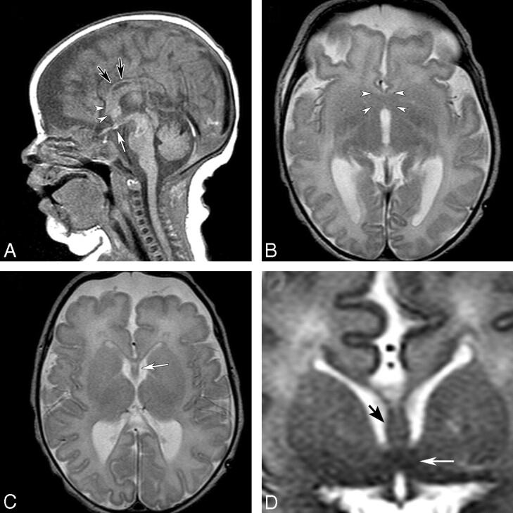
Fig 3.
MR imaging of an 8-day-old term neonate with atrial and ventricular septal defects and SMMCI. A, Sagittal T1-weighted image shows a thin CC (black arrows), a thickened fornix, and dysplastic subcallosal areas (arrowheads). A small ectopic pituitary gland is noted near the chiasm (white arrow). B, Axial T2-weighted image shows an area of fusion in the septal and preoptic regions (arrowheads). C, An azygous ACA is present in the anterior IHF. Axial T2-weighted image slightly superior shows the presence of the SP (white arrow) and a thin genu of the CC. D, Coronal T2-weighted image at the level of the AC shows fusion of the midline region (white arrow) below the fornices (black arrow).

