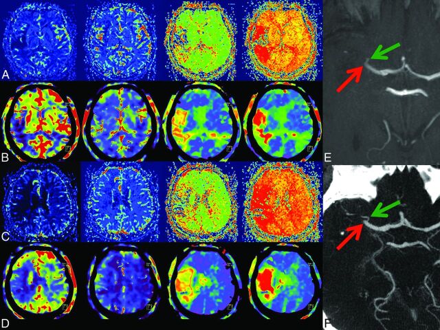
Fig 4.
2) Case example. In rows A and C, MRP; in B and D, corresponding FD-CTP parameters maps (left to right: CBF, CBV, MTT, and TTP) are displayed. A and B and C and D are corresponding MRP and FD-CTP images. Due to an MCA occlusion, there is a significant perfusion deficit in the CBF, MTT, and TTP maps. There is no significant CBV lesion in this patient. The MCA occlusion was visible in either the MRA (E, red arrow) or in FD-CTA (F, red arrow) MIP reconstruction. The green arrow indicates a small temporal branch.

