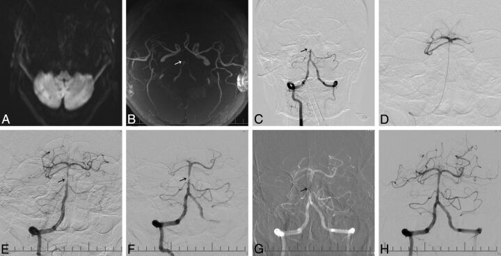
Fig 1.
A 54-year-old male patient who presented with vertigo and decreased consciousness for 10 hours. A, MR imaging shows multiple infarcts over the bilateral cerebellar hemispheres on DWI. B, MRA shows basilar artery occlusion (arrow). C, A right vertebral angiogram shows occlusion of the basilar artery (arrow). D, An angiogram after crossing of the basilar artery occlusion with a microcatheter shows patent distal flow at the basilar artery tip, with opacification of both posterior cerebral arteries and superior cerebellar arteries. E, Angiogram after deployment of the Solitaire AB device shows restoration of flow in the basilar artery, with suspected thrombus and focal stenosis (arrow) in the mid-distal segment of the basilar artery. F, Angiogram post-mechanical thrombectomy shows underlying severe focal stenosis of the basilar artery (arrow). G, Deployment of an Apollo stent (arrow) at the site of the basilar artery stenosis. H, Final angiography shows a TICI flow of grade 3 in the basilar artery with good distal perfusion.

