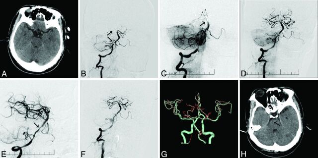
Fig 2.
A 55-year-old male patient who presented with tetraparesis and decreased consciousness for 6 hours. A, Plain CT on admission does not reveal any large territory infarct. B, The right vertebral angiography shows occlusion at the tip of the basilar artery (arrow) and right posterior cerebral artery and severe stenosis of the right vertebrobasilar junction (arrows). C, Angiography postdeployment of the Solitaire AB device at the proximal right posterior cerebral artery shows no opacification of the distal basilar artery (arrow). D, Angiography post-mechanical thrombectomy shows opacification of the distal basilar artery and both posterior cerebral arteries and severe stenosis in the vertebrobasilar junction. E, Deployment of a Wingspan stent (arrow) at the vertebrobasilar junction. F, Right vertebral angiography after stent placement shows resolution of the proximal basilar artery stenosis and good contrast flow in the basilar artery (TICI grade 3). G–H, Follow-up CT and CTA at 24 hours show a patent basilar artery and a small right pontine infarct.

