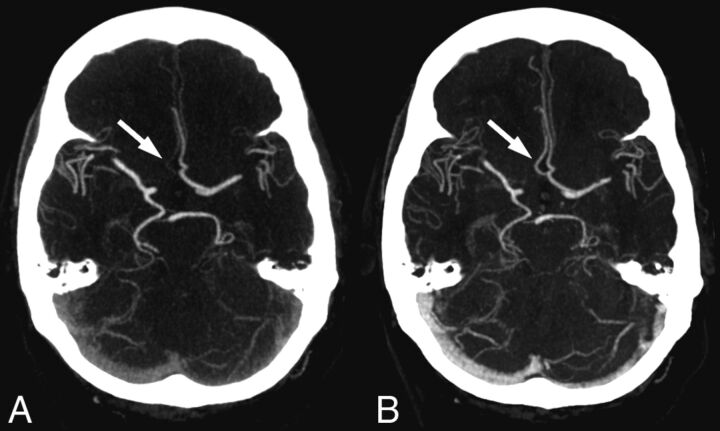
Fig 3.
Images show the effect of delayed contrast material arrival in a patient with right-sided hemiparesis. A, On standard CT angiography, the right-sided anterior cerebral artery was considered occluded in both segments A1 and A2. B, On timing-invariant CTA, the right-sided anterior cerebral artery was considered hypoplastic in segment A1 and patent in segment A2. Standard CTA shows only faint enhancement in the right-sided A2 segment (arrow) because the bulk of contrast material arrived after the standard CTA acquisition. Timing-invariant CT shows strong A2 enhancement (arrow) because it is delay-insensitive and displays maximal contrast enhancement with time.

