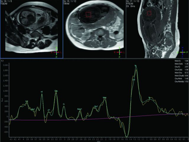
Fig 4.
GW 31 + 3, single-voxel MR spectroscopy of the brain. PRESS with a short TE (35 ms). VOI 2.7 cm3. Breech presentation. Peak in the lipid/Lac region. No fetal pathology was found. In this case, a premature rupture of the membranes was present and indications of maternal infection, which confirmed the decision to perform a cesarean delivery. Notice the reduced amniotic fluid on the scout scans.

