
Fig 3.
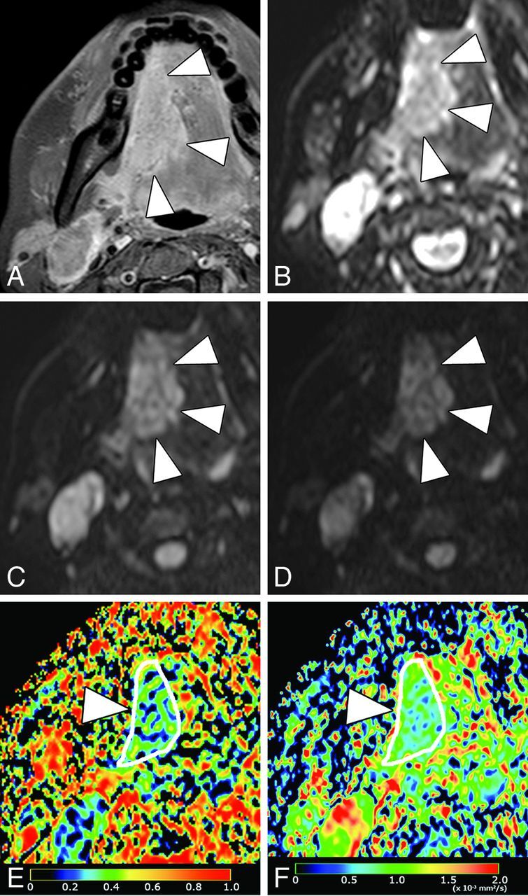
A 37-year-old woman with SCC in the tongue. A, Axial fat-suppressed contrast-enhanced T1-weighted image shows enhancing tumor (arrowheads), which occupies the right anterior two-thirds of tongue. B, Axial DWI at b = 0 s/mm2. C, Axial DWI at b = 500 s/mm2. D, Axial DWI at b = 1000 s/mm2. E, Axial PP map shows the tumor (arrowheads) with small perfusion (average PP value = 0.14). F, Axial D map shows the tumor (arrowhead) with small diffusion (average D value = 0.68 × 10⁻³ mm2/s).