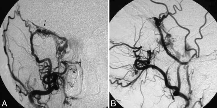
Fig 2.
Patient 8. Cerebral angiograms show a fistula (arrow) along the lesser sphenoid wing, primarily fed by the right MMA and draining into the right cavernous sinus, bilateral inferior petrosal sinuses, and the right superior ophthalmic vein. A, Anteroposterior view of the right ECA angiogram. B, Lateral view of the right ECA angiogram.

