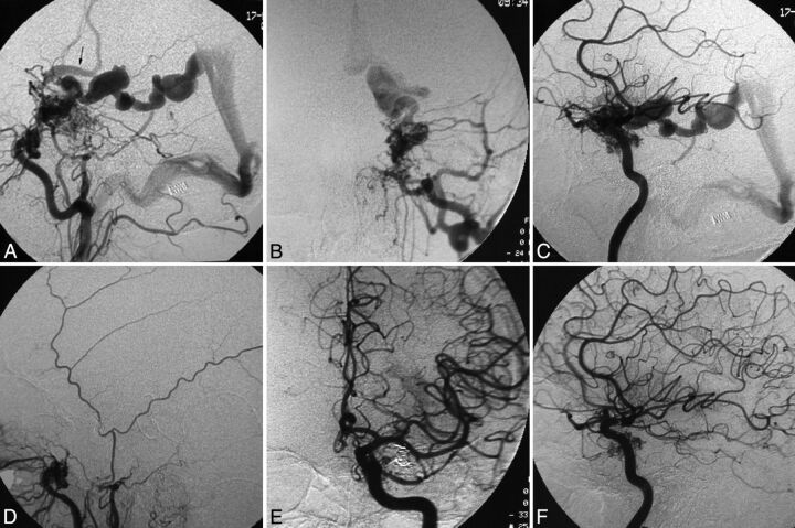
Fig 3.
Patient 5. The ECA (A, lateral view; B, anteroposterior view) and ICA (C, lateral view) angiograms show that the high-flow fistula is fed by multiple branches of the ICA and the ECA, and there is venous drainage into the basal vein of Rosenthal and subsequently the vein of Galen, as well as into the Sylvian and Trolard veins (arrow) into the superior sagittal sinus. A triaxial system with a guide catheter inserted into the internal jugular vein, an intervening Tracker-38 catheter (Boston Scientific) placed in the transverse sinus near the torcula—and, in this, a longer microcatheter was navigated to the exact fistula site through the straight sinus, the vein of Galen, and the basal vein of Rosenthal—and transvenous embolization of the fistula was performed using coils. Postembolization angiograms of the ECA (D, lateral view) and ICA (E, anteroposterior view; F, lateral view) show complete obliteration of the fistula.

