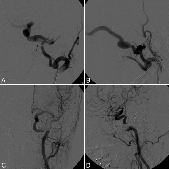
Fig 4.
Patient 3. Anteroposterior (A) and lateral (B) view of the left ECA angiograms during transarterial coil embolization show that the fistula site involves the sphenobasal sinus (arrow) draining to the left cavernous sinus and SOV. Five-month follow-up angiogram of the left common carotid artery (C, anteroposterior view; D, lateral view) demonstrates durable complete obliteration of the fistula by Onyx and coils.

