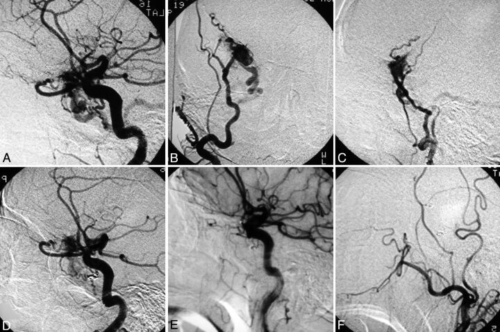
Fig 5.
Patient 11. Lateral angiographic view of the right ICA (A) shows primary feeding arteries from the recurrent meningeal branch of the ophthalmic artery as well as the inferolateral trunk of the ICA, with resultant venous drainage into an enlarged vein of Labbe. Anteroposterior (B) and lateral (C) view of the right internal maxillary artery angiogram showing a fistula at the lesser sphenoid wing draining into the right temporal lobe cortical vein. Lateral view of the ICA (D) after transarterial coiling and n-BCA injection of the fistula showing a residual lesion. Lateral view of the right ICA (E) and ECA (F) at 18-month follow-up show complete obliteration of the fistula.

